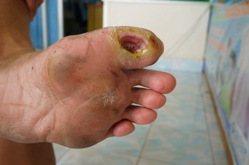
INJURY PREVENTION

Achilles Tendonitis

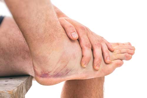
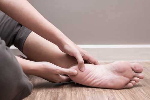
Achilles tendonitis is a painful and often debilitating inflammation of the Achilles tendon (heel cord). The Achilles tendon is the largest and strongest tendon in the body. It is located in the back of the lower leg, attached to the heel bone (calcaneus), and connects the leg muscles to the foot.

The Achilles tendon gives us the ability to rise up on our toes, facilitating the act of walking, and Achilles tendonitis can make walking difficult almost impossible.

There are the three stages of tendon inflammation:

Achilles tendonitis that occurs as a result of arthritis in the heel is more common in people who are middle aged and older.

Poorly conditioned athletes are at the highest risk of developing Achilles tendonitis. Participating in activities that involve sudden stops and starts and repetitive jumping (e.g. basketball, tennis, dancing) increased the risk for the condition. It often develops following sudden changes in activity level, training on poor surfaces, or wearing inappropriate footwear.

Achilles tendonitis may be caused by a single incident of overstressing the tendon, or it may result from a series of stresses that produce small tears over time (overuse). Patients who develop arthritis in the heel have an increased risk for developing Achilles tendonitis. This occurs more often in people who are middle aged and older.

Download Information Sheet (PDF)

Ankle Sprain

A sprain is a stretched or torn ligament. Ligaments connect one bone to another bone at a joint and help keep the bones from moving out of place.

The most common site of sprains is the ankle. An ankle sprain can happen when you fall, when you suddenly twist your ankle too far, or when you force the joint out of its normal position.

Most ankle sprains occur during sports activities or when walking or running on an uneven surface.

The signs of an ankle sprain can include:

The severity of an ankle sprain depends on how badly the ligaments are stretched or torn. If the sprain is mild, there may not be much pain or swelling, and the ligaments may only be stretched. If sprain is severe, one or more ligaments may be torn, and the joint may be severely swollen. A severe sprain can also be extremely painful.

Rest: You may need to rest your ankle

Ice: Using ice packs

Compression: Wrapping your ankle may be the best way to avoid swelling and bruising

Elevation: Raising your ankle to or above the level of your heart will prevent the swelling from getting worse and will help reduce bruising. Try to keep your ankle elevated for about 2 to 3 hours a day if possible.

Arthritis

Osteoarthritis is the broad term used to describe degenerative joint disease, it is a condition which the hyaline cartilage that lines the surface of the bones forming a joint are eroded or damaged. As the cartilage wears inflammation and pain occurs, the sufferer generally experiences pain upon weight-bearing and walking, ultimately arthritis results in decreased joint range of motion and therefore muscle atrophy.

Arthritis in one or more joints of the foot will alter the normal biomechanics of walking and therefore make walking difficult.

The main causes of osteoarthritis of the foot are:

Arthritis in the feet usually presents with one or more of the following symptoms: joint swelling and tenderness, redness or heat, limited motion and stiffness, and pain when walking. Common associated foot conditions are bunions, hammer toes, claw toes and corns and callus resulting from swollen joints.
Arthritis involves evaluation of signs and symptoms, the use of x-ray, CT scan and blood tests may be of use to determine the severity, type and location. Management of arthritis usually resolves around maintain an acceptable level of comfort and mobility to the sufferer. The use of medications such as analgesics and non-steroidal anti-inflammatory drugs are standard practice, gentle exercise to maintain muscle tone and joint range of motion are advised. Appropriate footwear and the use of innersoles or orthotics may be beneficial; in severe cases surgery may be required.

Athlete's Foot (tinea)

Athlete’s foot is a common infection caused by the tinea fungus. It is not serious. Symptoms include itching, burning and cracked, scaly skin between your toes. Tinea grows best in damp, dark and warm places, which is why it often develops between your toes. It can spread to your toenails, as well, making them thick and crumbly.

Athlete’s foot causes scaling, faking and itching of the affected skin. Blisters and cracked skin may also occur, leading to exposed raw tissue, pain, swelling and inflammation. Secondary bacterial infection can accompany the fungal infection, sometimes requiring a course of oral antibiotics.

The infection can be spread to other areas of the body, such as the groin, and usually is called by a different name once it spreads, such as tinea corporis on the body or limbs and tinea cruris for an infection of the groin. Tinea pedis most often manifests between the toes, with the web space between the fourth and fifth digits most commonly afflicted.

You can get athletes foot from damp surfaces, such as locker room floors. To prevent it:

Treatments include over-the-counter antifungal creams for most cases and prescription medicines for more serious infections.

Blisters

Blisters are small swellings of the skin that contains watery fluid, usually caused by friction, the body responds to friction by producing fluid, the fluid builds up beneath the part of the skin being rubbed, causing pressure and pain. Blisters are a common problem with athletes wearing new shoes, or during extended periods of walking or running, as friction and heat are generated.
If the blister has not been broken leave it alone, most blisters are reabsorbed and heat on their own, deflective padding or cushioned innersoles may ease symptoms and prevent recurrence of blister. If the blister has broken, flush the area with saline solution and dress with band aid, antiseptics may help prevent infection.

Callus and Corns

A callus is an especially toughened area of skin which has become relatively thick and hard as a response to repeated contact or pressure. A corn is a specially-shaped callus of dead skin that usually occurs on thin or glabrous skin surfaces, especially on the dorsa of toes or fingers.
Corns appear as a horny thickening of the skin on toes. This thickening appears as a cone shaped mass pointing down into the skin. Hard corns are usually located on the outer surface of the little toe or on the upper surface of the other toes, but can occur between the toes. A soft corn occurs between the toes and is kept soft by the moisture in this area. A callus is more diffuse area of thickening and does not have the focal point of the corn.
Corns and callus are caused by one thing – TOO MUCH PRESSURE, usually in combination with some friction. There is no other way to get them; the pressure stimulates the skin to thicken to protect itself, but as the stimulation of the pressure continues, it becomes painful.
Self treatment or management of corns and callus includes:
  • following the advice of a Podiatrist
  • proper fitting of footwear
  • proper foot hygiene and the use of emollients to keep the skin in good condition

Claw Toes

Claw toes are classified on the mobility of the joints. There are two types – flexible and rigid. In a flexible claw toe, the joint has the ability to move. This type of claw toe can be straightened manually. A rigid claw toe does not have that same mobility to move. Movement is very limited and can be extremely painful. This sometimes causes foot movement to become restricted leading to extra stress at the ball of the foot, and possible causing pain and the development of corns and calluses.
  • Your toes are bent upward from the joints at the ball of the foot.
  • Your toes are bent forward at the middle joints toward the sole of your shoe
  • Sometimes your toes also bend downward at the top joints, curling under the foot.
  • Corns may develop over the top of the toe or under the ball of the foot.
  • Wear shoes with soft, roomy toe boxes and avoid tight shoes and high-heels.
  • Use your hands to stretch your toes and toe joints toward their normal positions.
  • Exercise your toes by using them to pick up marbles or crumple a towel laid flat on the floor.
  • A special pad can redistribute your weight and relieve pressure on the ball of the foot.
  • Ask a shoe repair shop to stretch a small pocket in the toe box.

Diabetic Ulcers

Foot problems are a big risk. Like all diabetic people, you should monitor your feet. If you don’t, the consequence can be severe, including amputation, or worse.

Minor injuries become major emergencies before you know it. With a diabetic foot, a wound as small as a blister from wearing a shoe that’s too tight can cause a lot of damage. Diabetes decreases your blood flow, so your injuries are slow to heal. When your wound is not healing, it’s at risk of infection. As a diabetic, your infections spread quickly.
 
  • Wash your feet everyday with mild soap.
  • When drying them, pat each foot with a towel and be careful between your toes.
  • Use quality lotion to keep the skin of your feet soft and moist – don’t put any lotion between your toes.
  • Trim your toe nails straight across. Avoid cutting the corners. Use a nail file or emery board. If you find an ingrown toenail, see your doctor.
  • Always keep your feet warm. Don’t get your feet wet in snow or rain.
  • Don’t smoke or sit cross-legged. Both decrease blood supply to your feet.
  • Never walk barefoot or in sandals or thongs.
  • Buy new shoes late in the day when your feet are larger. Buy shoes that are comfortable without a “breaking in” period. Try to get shoes made with leather upper material and deep toe boxes.
  • Avoid socks with holes or wrinkles and dry socks everyday.
  • Square toed socks will not squeeze your toes. Avoid stockings with elastic tops.

Genu Valgum (knock knees)

 In knock knees, the lower legs are at an outward angle, such that when the knees are touching, the ankles are separated.

Most children develop a slight knock-kneed stance by the time they are 2 or 3 years old, often with significant separation at the ankles when the knees are touching. This is part of normal development and often persists through age 5 or 6, at which time the legs begin to straighten fully. By puberty, most children can stand with the knees and ankles touching (without forcing the position). Knock knees can also develop as a result of disease processes. Most often the cause has already been diagnosed and the knock knees are recognized as a symptom of the condition.

Generally, there is no known cure for knock knees post-childhood. Contrary to common belief, no amount of orthotic treatment or bodybuilding exercise will straighten knock knees for adults. If the condition persists or worsens into late childhood and adulthood, a corrective osteotomy may be recommended to straighten the legs. This however is more of a cosmetic remedy, and may hamper athletic performance in the future.

Adults with uncorrected genu valgum are typically prone to injury and chronic knee problems such as chondromalacia and osteoarthritis. In some cases, total knee replacement (TKR) surgery may be required later in life to relieve pain and complications resulting from severe genu valgum.

Genu Varum (bow legs)

Genu Varum is a deformity marked by medial angulations of the leg in relation to the thigh, an outward bowing of the legs, giving the appearance of a bow. It is also known as bandy-leg, bowleg, and bow-leg and tibia vara. Usually there is an outward curvature of both femur and tibia.

If a child is sickly, wither with rickets or suffering from any ailment that prevents the due ossification of the bones, or is improperly fed, the bowed condition may remain persistent. Thus the chief cause of this deformity is rickets. Skeletal problems, infection, and tumors can also affect the growth of the leg, sometimes giving rise to a one-sided bow-leggedness. The remaining causes are occupational, especially among jockeys, and from physical trauma, the condition being very likely to supervene after accidents involving the condyles of the femur.

Generally, no treatment is required for idiopathic presentation as it is a normal anatomical variant in young children. Treatment is indicated when it persists beyond 3 and a half years old, unilateral presentation or progressive worsening of the curvature. When caused by rickets, the most important thing is to treat the constitutional disease, at the same time instructing the mother never to place the child on its feet. In many cases this is quite sufficient in itself to affect a cure, but matters can be hastened somewhat by applying splints. When the deformity arises in older patients, either from trauma or occupation, the only treatment is surgery.

Gout

Gout is a painful arthritic condition caused by uric acid crystals forming in the joint capsule.

Uric acid is generated as the body’s tissues are broke down during normal cell regeneration, some people with gout generate too much uric acid (10%) or more commonly (90%) fail to effectively eliminate their uric acid into the urine. Genetics and nutrition seem to be the key factors in the development of gout, however trauma and medications can be contributing causes.

Gout symptoms include severe pain and tenderness, redness, heat and swelling in the affected joints, most commonly in the big toe or metatarso-phalangeal joint.

Commonly the pain and other symptoms of gout can be managed by non-steroidal anti-inflammatory drugs (NSAIDS), the use of medications such as Allupurinol may help reduce serum uric acid levels and prevent further attacks of gout. Also dietary and lifestyle changes including reduction of alcohol and red meat are recommended.

Hallux Valgus (bunions)

 A bunion (hallux Valgus) is a structural deformity of the bones and the joint between the foot and big toe, and may be painful.

A bunion is an enlargement of bone or tissue around the joint at the base of the big toe (metatarsophalangeal joint). The big toe may turn in toward the second toe (angulation), and the tissues surrounding the joint may be swollen and tender.

Bunions may be treated conservatively with changes in shoe gear, different orthotics (accommodative padding and shielding), rest, ice, and medications. These sorts of treatments address symptoms more than they correct the actual deformity. Surgery, by a podiatrist, may be necessary if discomfort is severe enough or when correction of the deformity is desired.

Commonly the pain and other symptoms of gout can be managed by non-steroidal anti-inflammatory drugs (NSAIDS), the use of medications such as Allupurinol may help reduce serum uric acid levels and prevent further attacks of gout. Also dietary and lifestyle changes including reduction of alcohol and red meat are recommended.

Orthotics are bunion cushions, splints, regulators while conservative measures include various footwear like gelled toe spacers, bunion/toe separators, bunion regulators, bunion splints, and bunion cushions.

Procedures are designed and chosen to correct a variety of pathologies that may be associated with the bunion. The age, health, lifestyle and activity level of the patient may also play a role in the choice of procedure.

Hammer and Mallet Toe

A hammer toe is a deformity of the middle joint of the second, third or fourth toe causing it to be permanently bent, resembling a hammer.

Hammer toe most frequently results from wearing poorly-fit shoes that can force the toe into a bent position. Such as excessively high heels or shoes that are too short or narrow for the foot. Having the toes bent for long periods of time can cause the muscles in them to shorten, resulting in the hammer toe deformity. This is often found in conjunction with bunions or other foot problems. It can also be caused by muscle, nerve, or joint damage resulting from conditions such as osteoarthritis, rheumatoid arthritis, Charcot-Marie-Tooth disease or diabetes.

In many cases, conservative treatment consisting of physical therapy and new shoes with soft, spacious toe boxes is enough to resolve the condition, while in more severe or longstanding cases orthopaedic surgery may be necessary to correct the deformity.

The patient’s doctor may also prescribe some toe exercises that can be done at home to stretch and strengthen the muscles. For example, the individual can gently stretch the toes manually, or use the toes to pick things up off the floor. While watching television or reading, he can put a towel flat under the feet and use the toes to crumple it.

Heel Spurs

Heel Spurs are often the cause of serious pain in the heel of the foot.The Spurs can easily interfere and disrupt your daily lifestyle as they generate aching pain with every step. Many factors can contribute to the development of a spur, but luckily there are conservative ways to stop pain and reverse the condition.

Pain that results from a heel spur is usually characterized by a sharp poking and is localized to the heel or under the heel. Aching pain can become very severe and inflammation and bruising can also occur. When taking a step or when putting pressure on the heel, pain can escalate, particularly after prolonged periods of rest like the first step after getting out of bed in the morning.

Most cases of heel spurs can be treated by the simple, conservative means of heel cups, heel seats, heel pads, arch supports, and innersoles. They use support/pressure as an effective way to treat the pain associated with heel spurs. The unique treatments not only effectively treat the pain; they also reverse the heel spur growth. They cradle the heel and arch of the foot, providing the support and reinforcement of the heel which is necessary to relieve stress and pain in the tissue around the heel spur.

Surgery, which is a more radical treatment, can be a permanent correction to remove the spur itself. If your doctor believes that surgery is indicated, they will recommend an operation – but only after establishing that less drastic methods of treatment are not successful.

Morton's Neuroma

This condition develops when one of the nerves running along the sole of the foot becomes squeezed, or inflamed between two of the bones of the foot (metatarsals).

Usually, related to poor shoe wear, or increase in running/standing. When the toes and foot are unnaturally squeezed, there is pressure created on the nerves in the foot. The nerve becomes chronically thickened and inflamed.

Pain in the ball of the foot, often described as burning, or numbness, that may radiate into the toes. The space between the third and forth toes is most common, but may occur in the space between the second and third toes as well. Tenderness, pain or numbness in the third or forth web space with direct pressure on the nerve.

Diagnosis is based almost entirely on history and physical examination. X-rays are usually normal. Ultrasound or MRI scans are the most useful. Ruling out other causes of foot pain is required.

Modification of shoes, allowing for a wider toe box and good arch and metatarsal support are important. Orthotics may be helpful with the addition of a metatarsal dome. Non-steroidal anti-inflammatory medication and rest are also advised. A trial of a corticosteroid injection may be of both diagnostic and therapeutic benefit.

Excision of the swollen nerve (neuroma) is generally curative; also mild numbness between the involved toes may result.

Metatarsalgia (ball of foot pain)

Metatarsalgia, a form of neuralgia, is an inflammation of the nerve that runs between the third and fourth metatarsal (foot) bones.

Metatarsalgia really covers a group of foot disorders. The classic symptom is pain in the ball of the foot. Many people say that it is “like walking on pebbles”, but x-rays usually show nothing irregular.

The problem affects males and females from adolescents to older adults. It is most common in middle-aged women who often wear high heels.

The most common causes are:

  • Heredity: Narrow, high-arched feet can focus stress on the balls of the feet.
  • Skin Irritation: Metatarsalgia often occurs with bunions or tender calluses under the metatarsal-phalangeal joints.
  • “Overloaded” Feet: Excess weight from pregnancy or obesity can contribute to Metatarsalgia.
  • Nerve Disorders: Morton’s Neuroma is a benign growth that can develop on a nerve in the foot.

In most cases, simple measures will lessen pain at the front of the foot.

  • Foot Freedom: Don’t wear tight shoes and high heels
  • Orthotics (or metatarsal pads): Consult your podiatrist or physician for a footpad that relieves pressure on the metatarsal area.
  • Medications: Your doctor may prescribe non-steroidal anti-inflammatory medications such as ibuprofen or sulindac. This is the most common treatment.
  • Surgery: An operation seldom is necessary.

Patello Femoral Syndrome (knee pain)

Patello-femoral syndrome or “runner’s knee” is one of the most common knee conditions affecting athletes. It can be defined as retro patella or peri-patella pain. It results from the kneecap (patella) not tracking smoothly through the groove in the underlying femoral bone when the leg is bent and straightened. Pain typically presents in the front of the knee, in and around the kneecap especially when going up or down stairs, squatting, kneeling or running.

Many factors may contribute to cause the kneecap to mal-track. Commonly, weakness of the inner quadriceps muscle plus poor lower limb biomechanics, over-pronation (feet rolling inwards), or overuse may result in the kneecap being pulled towards the outside of the leg.

Initial treatment suggested is typical for almost all acute sporting injuries. Icing the knee for 10-20 minutes after activity should help reduce inflammation and pain, plus reducing or resting from the activity that causes the pain. The use of simple painkilling medication such as paracetamol and anti-inflammatories may help.

  • Taping or strapping the kneecap may be of some benefit (see your Podiatrist or Physiotherapist for correct strapping technique)
  • Correction of lower limb biomechanics, overpronation via the use of a Realign innersole or prescription orthotics prescribed by Podiatrist, plus muscle stretching and strengthening exercises is often beneficial (again see your Podiatrist or Physiotherapist for the appropriate exercise program
  • Appropriate sport specific footwear with adequate support to enhance function and assist in addressing any biomechanical anomalies

Pes Cavus (high arches)

Pes Cavus is an excessively raised medial arch (also called instep) on the bottom of the foot. The arch runs from the toes to the heel on the inside part of the foot.

The symptoms of a high arch foot will vary depending on how severe the condition is and the activity levels of the person with it. Most will have no pain or any other symptoms. Symptoms may vary from a mild problem with shoe fitting to significant disability.

Unlike flat feet, highly arched feet tend to be painful because more stress is placed on the section of the foot between the ankle and the toes (metatarsal). This condition generally makes it difficult to fit shoes. In addition, those with high arches usually need foot support.

The symptoms of a high arch foot will vary depending on how severe the condition is and the activity levels of the person with it. Most will have no pain or any other symptoms. Symptoms may vary from a mild problem with shoe fitting to significant disability.

  • There may be corns and calluses under the bases of the first and fifth toes.
  • Shoe may not fit very well because of the high arch and the clawed toes.
  • There may be some pain in the arch area, because of the pressure that it is under.
  • The feet will feel stiffer and less mobile than a foot that does not have a high arch.
  • An ankle sprain is more common in those with a high arched foot.

Pes Planus (flat feet)

Pes Planus is a condition where the arch or instep of the foot collapses and comes in contact with the ground. In some individuals, this arch never develops.

Flat feet are a common condition. In infants and toddlers, the longitudinal arch is not developed and flat feet are normal. The arch develops in childhood, and by adulthood, most people have developed normal arches.

When flat feet persist, the majority are considered variations of normal. Most feet are flexible and an arch appears when the person stands on his or her toes. Stiff, inflexible or painful flat feet may be associated with other conditions and require attention.

Painful flat feet in children may be caused by a condition called tarsal coalition. In tarsal coalition, two or more of the bones in the foot fuse together, limiting motion and often leading to a flat foot. Most flat feet do not cause pain or other problems.

Flat feet may be associated with pronation, a leaning inward of the ankle bones toward the center line. Shoes of children who pronate, when placed side by side, will lean toward each other (after they have been worn long enough for the foot position to remodel their shape).

Foot pain, ankle pain, or lower leg pain (especially in children) may be a result of flat feet and should be evaluated by a health care provider.

  • Absence of longitudinal arch of foot when standing
  • Generalised foot pain
  • Heel tilts away from the midline of the body more than usual.

Plantarfasciitis (arch pain)

Plantar fasciitis is irritation and swelling of the thick tissue in the bottom of the foot.

The plantar fascia is a very thick band of tissue that covers the bones on the bottom of the foot. This fascia can become inflamed and painful in some people, making walking more difficult. Risk factors for plantar fasciitis include foot arch problems (both flat foot and high arches), obesity, sudden weight gain, running, and a tight Achilles tendon (the tendon connecting the calf muscles to the heel).

This condition is one of the most common complaints relating to the foot. Plantar fasciitis is commonly thought of as being caused by a heel spur, but researched has found that this is not always the case.

On x-ray, heel spurs are seen commonly both in people with and without plantar fasciitis

The most common complaint is pain in the bottom of the heel, usually worst in the morning and improving throughout the day. By the end of the day the pain may be replaced by a dull aching that improves with rest.

  • Losing weight.
  • Ice after activity. Plain ice is good, and rest
  • Stretching the calf muscles several times a day.
  • Arch support innersoles and Orthotics.
  • Supportive footwear.

Shin Splints

Shin Splints (medial tibial stress syndrome) is the general term used to describe inflammation of the postero-medial and anterior crest of the tibia, generally associated with overuse of the soleus fascia or the peri-osteal tissue beneath the posterior tibial muscle. It is a slow healing and painful condition and it is anticipated that between 10-15% of running injuries are shin splints.

Commonly shin splints occur due to overuse or at the beginning of an exercise program if commenced too aggressively. There are often certain underlying biomechanical factors contributing to its development. Excessive pronation and poor shock attenuation may enhance eccentric contractions of the leg muscles and contribute to the development of shin splints. Shin splints can be classified into four grades.

  • GRADE 1: Pain on palpation of the medial tibial crest – asymptomatic when running
  • GRADE 2: Pain or discomfort after activity – but not during running
  • GRADE 3: Pain when running and residual discomfort after activity
  • GRADE 4: Pain and discomfort when engaged in simple walking
  • Early conservative treatment usually leads to a successful outcome. All sporting activity should be ceased or significantly reduced. Icing the painful shin region, immobilisation and if necessary anti-inflammatory medication may help.
  • A physiotherapy assessment and program incorporating deep tissue massage, ultrasound and stretching and strengthening may also be required. This should also be continued on cessation of symptoms to prevent recurrence of original symptoms.
  • The recognition and attention to any biomechanical abnormalities should be addressed with appropriate Realign innersoles or a visit to a Podiatrist for specialised biomechanical assessment and prescription orthotics may be helpful.
  • Appropriate supportive footwear may be of some benefit.

Tibialis Posterior Dysfunction

Tibialis Posterior Dysfunction is a progressive inability of the TIbialis Posterior tendon to control subtaler joint pronation, often resulting in acquired flat foot deformity and pain.

There are four recognized types of Tibialis Posterior Dysfunction:

  • Type I – Caused by direct injury such as laceration
  • Type II – Rupture secondary to other disease processes – ie. Rheumatoid arthritis and gout.
  • Type III – Idiopathic, unknown cause
  • Type IV – Functional, secondary to long term biomechanical forces ie. Excessive subtaler joint pronation

Initial conservative treatment should include rest, oral Non Steroidal Anti-Inflammatory Drugs (NSAIDS) and physical therapy. The use of orthotics to support the medial arch is recommended.

When conservative treatment fails, surgical intervention may be required.

Select the fields to be shown. Others will be hidden. Drag and drop to rearrange the order.
  • Image
  • SKU
  • Rating
  • Price
  • Stock
  • Availability
  • Add to cart
  • Description
  • Content
  • Weight
  • Dimensions
  • Additional information
Click outside to hide the comparison bar
Compare